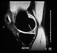

The evolution of MR angiography (MRA), CT angiography (CTA), and ultrasound angiography techniques have been significant developments, and they are replacing x-ray angiography as the preferred diagnostic tool for the detection of plaques and blockages (stenoses) in the blood vessels. This is due to the fact that conventional angiography is more invasive, significantly more time consuming, more expensive than MRA, CTA and ultrasound angiography. Thus, conventional angiography is being increasingly used only as a therapeutic tool to aid, for instance, in angioplasty procedures, which are helpful in removing plaque obstructions in the neck vessels or for opening clogged coronary arteries.

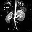
MR Angiogram of the descending aorta, kidneys and renal arteries
MR is also developing a tremendous potential for not only showing the structure or anatomy of the body, but also the function or workings of the body. For instance, some leading MR companies have commercially available systems (with echo Planar Imaging or EPI capabilities) that can show brain function, thus making the early detection of deadly stroke more possible. Functional MR (f-MR) can be used to even glimpse at the neural activity of the brain itself, for example, during pattern recognition study. f-MR can also show the function of the cardiac muscle (myocardium). Other MR procedures can depict the coronary vessels or create movie-like scenes of the beating heart. While these functional studies are not yet commonplace, even at many large university medical centers, use of these functional studies and their promising benefits may proliferate in the coming years as they become easier and less expensive to perform.
MRI, like CT, requires the patient to lie still on a special table inside the scanning gantry for ten to thirty minutes. However, during the conventional "high-field" MR exam, patients must lie completely still for shorter durations of five t o ten minutes at a time. The CT exam may only require patient cooperation for a few seconds to a few minutes at a time. Due to the longer examination times of MR and the closed nature of the cylindrical magnet used in traditional "high-field" MR equipment (0.5 to 1.5 Tesla magnetic field strength), some patients who undergo MR may experience claustrophobic anxiety.
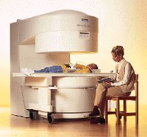
A mother reads to her daughter during an Open MR examination
Technical innovation is bringing new, "friendlier" MR designs like "open MR" and "short-bore MR", into routine clinical use. The new "open" or "C-shaped" MR systems (0.2 to 0.3 Tesla field strength) are typically open on all sides and improve a person's MR experience by lessening claustrophobia. Also becoming available are new versions of conventional "high-field" (cylindrical) MR systems with shorter magnets and more patient friendly (wider and shorter) aperture designs. These new "short-bore" systems promise to also further minimize patient anxiety about MR examination.
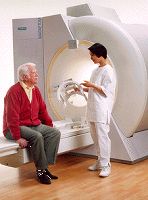
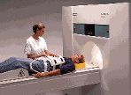
The MR technologist consoles a senior prior to an MR examination on a new "short bore system"


